Advanced Dental Technology
 At Danville Dental Associates, our skilled dentists and team proudly incorporate the most innovative dental techniques and technologies in order to make patient care faster, more effective, and more comfortable. From precise diagnostics to advanced treatment planning, our Danville and Chatham dental practice locations use dental technology to improve every aspect of the patient experience. If you’d like to find out more about our office or schedule an appointment, give our caring team a call. We welcome patients from Chatham, Ringgold, and other nearby communities.
Intraoral Camera
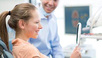
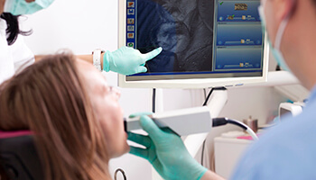 You’ve probably heard the saying, “a picture is worth a thousand words.” You’ve probably also heard your dentist struggling to explain a complex oral health condition to you while using tiny mirrors to show you a blurred image of your smile. That’s why we use intraoral cameras to capture crystal clear photos of your teeth, gums, and other oral structures. We display these images on chairside computer monitors, and using these pictures, we can more easily explain your potential oral health concerns and treatment options.
Digital X-Rays
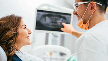 Like digital images that allow us to see the surface-level parts of teeth, digital x-rays give our team a view of the internal structures of teeth. Digital x-rays are faster and easier to capture than traditional, film radiography. Traditional x-rays required hazardous chemicals to develop and had to be viewed on a special light board. They were difficult to store and transfer physically, and they produced images of a much lower quality than those possible with digital x-rays. Conversely, digital images are quick and comfortable to capture, and these high-definition images are immediately ready for view on chairside computer monitors. The electronic files are easily stored from year to year and can be instantly transferred to labs and specialists as needed.
CT/Conebeam Scanner
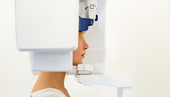 Digital x-rays give us clear images of specific parts of the smile. However, for more advanced treatment planning and diagnostics, it’s important that we have a picture of how the entire smile fits together. That’s where our state-of-the-art CT/Conebeam scanner comes into play. This scanner allows us to gather a full, panoramic image of the entire smile. We use CT imaging as part of the diagnostic process, especially in younger patients. It allows us to determine at the earliest stages whether or not your child’s smile is developing in proper alignment. We also use these scans as part of orthodontic treatment planning, so our dentist is able to accurately plan to reposition the bite into its ideal alignment.
Digital Impression System
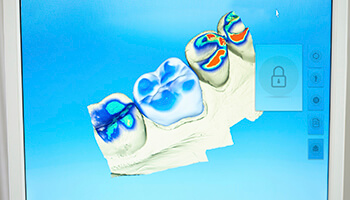 Traditional impressions are an essential part of dental practice. By biting into a malleable putty, patients leave behind a reflection of their bite. This reflected impression is used by dental labs to craft dental prosthetics like crowns, cosmetic solutions like porcelain veneers, and as part of the treatment planning process for orthodontics. Unfortunately, these impressions had to be physically shipped to labs, increasing the amount of time between the oral health care planning process and the implementation of treatments. Digital impressions offer a variety of benefits compared with traditional bite impressions including:
- Increased precision in capturing minute pits and grooves in teeth
- More comfortable to capture since patients will not need to bite into putty material
- Reduced time from planning to implementation of treatment since digital impressions are instantly transferable to our lab
We use digital impressions to improve our Invisalign® orthodontic treatments. We’re able to more accurately plan each set of alignment trays using digital impressions, and these more precisely designed aligners are crafted more quickly since the lab receives our impressions immediately.
Soft Tissue Laser
 When you think of lasers you might think that they’re just for SyFy films, but medical and dental professionals have been using laser technology to improve the quality and comfort of a variety of treatments for decades. Laser is actually an acronym that stands for light amplification by stimulated emission of radiation. Sounds complicated, but really that just means that lasers allow us to amplify or increase the amount of light using radiation, similar to that released during x-rays. These amplified light waves allow us to remove or reshape soft tissue with minimal bleeding and discomfort during treatment. Best of all, healing time is significantly reduced, and the risk of infection is negligible, which is essential in the laser treatment of gum disease.